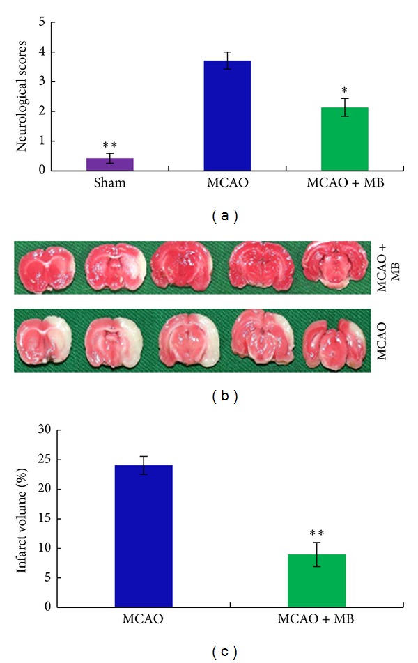
Figure 2.

The comparison on the protective effects of treatments on the neurological deficits and the infarct volume after ischemia stroke. The rats were administrated intraperitoneally by the tested formula drug at 30 min before ischemia and at 2 h, 12 h, and 24 h after ischemia. After 2 h of ischemia and 24 h of reperfusion, the neurological scores were evaluated according to a graded scoring system described in the method in Section 2.3, and the infarct volume was calculated by the method described in Section 2.4. (a) Represented the different neurological deficits scores of sham group, MCAO group, and formula-treated group; (b) was the representative images of TTC-staining brains from MCAO group and the formula-treated group; the normal brain tissue displayed the color of rose red; meanwhile the infarct area displayed a color of pale white; (c) showed the different infarct volumes from rats of sham group, MCAO group, and formula-treated group. Bars represent means ± SEM of six rats; *significant difference at P≦0.05. **Remarkably significant difference at P≦0.01.
