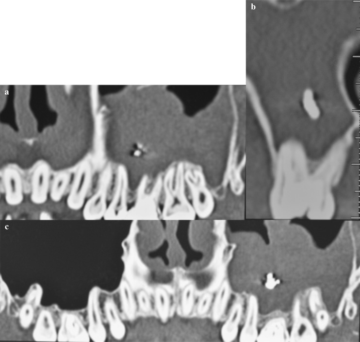
Figure 2 a, b, c.
(a) Image TC Dentascan shows a tissue with the density of the soft tissues located at the level of the left maxillary sinus, with formations likely calcic nature. (b) Paraxial image in correspondence of 2.6 in which it is possible to appreciate the root canal and calcification in the context of the maxillary sinus that appears obliterated at that level. (c) Image TC Dentascan shows the normal pneumatization of the right maxillary sinus.

