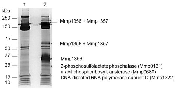
Fig. 4.
Pull-down of Mmp1356 expressed in M. maripaludis. The proteins purified by metal affinity chromatography from M. maripaludis cells transformed with the empty vector (lane 1) or the shuttle vector expressing His6-tagged Mmp1356 (lane 2) were analyzed with SDS-PAGE and stained with silver. The protein bands marked with arrows in lane 2 were excised, digested with trypsin, and analyzed by LC-MS/MS. The corresponding regions in lane 1 were analyzed as controls. The proteins specifically identified in lane 2 with > 40% coverage are listed.

