
Figure 1.
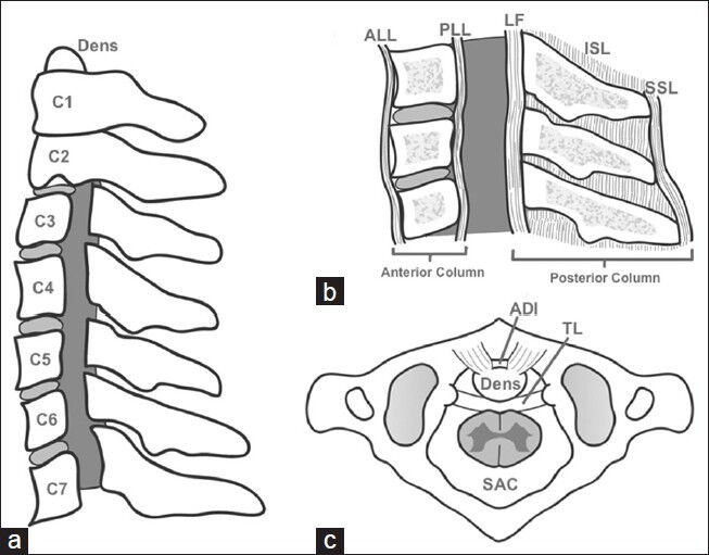
Cervical Spine Anatomy. (a) Lateral view of seven cervical vertebra, laminae and pedicles removed to show spinal cord space (gray), (b) Ligaments forming the anterior and posterior columns shown in lateral cross section, ALL: Anterior longitudinal ligament, PLL: Posterior longitudinal ligament, LF: Ligamentum flavum, ISL: Interspinous ligament, SSL: Supraspinous ligament, (c) Superior view of first on second cervical vertebra and the transverse ligament (TL), which normally limits translation and the atlas-dens interval (ADI)