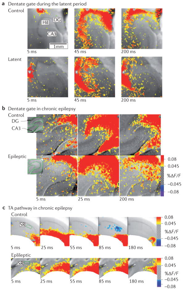
Figure 4. Circuit dysfunction in temporal lobe epilepsy.
a | Dentate gating is impaired during the latent period (1 week after status epilepticus) in an animal model of chronic temporal lobe epilepsy (TLE). The top panel shows voltage-sensitive dye (VSD) recordings of perforant path stimulation under control conditions 5 ms after stimulation (left), at peak (middle) and later (200 ms; right). The top right panel shows restriction of the perforant path-evoked depolarization to the molecular layer of the dentate gyrus (DG). Note that there is minimal area CA3 activation. During the latent period (bottom panel), identical stimulation reveals a delayed peak response as well as >200% increase in activation (as measured by areal pixel activation) in area CA3. b | Gating function is retained in the chronic phase of acquired TLE in the rodent model. The two panels show VSD images of the DG 5 ms (left), 25 ms (middle) and 200 ms (right) after stimulation of the perforant path in control rats (top panel) and in chronically epileptic rats (bottom panel). Under both conditions, there is a strong response in the DG that fails to propagate to area CA3. c | Temporoammonic (TA) pathway dysfunction in the chronic phase of acquired TLE. VSD imaging of TA pathway function in control animals (top panel) and epileptic animals (bottom panel) at 5 ms, 25 ms, 55 ms, 85 ms and 180 ms after stimulation of the TA pathway, producing spatially restricted activity in control animals that aberrantly propagates to the strata radiatum and pyramidale in the epileptic condition. Hil, hilus; F, fluorescence. Part a is reproduced, with permission, from REF. 148 © (2007) Society for Neuroscience. Parts b and c are reproduced, with permission, from REF. 144 © (2006) Society for Neuroscience.

