
Figure 3.
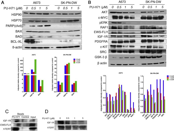
PU‐H71 induces production of co‐chaperone protein, HSP70. Pro‐apoptotic proteins are increased and anti apoptotic proteins are decreased in Ewing cell lines treated with PU‐H71. A, immunoblot analysis of the indicated proteins in A673 and SK‐PN‐DW cell lines treated with 0, 0.5, 1, 5 μM concentrations of PU‐H71 for 24 h. B, immunoblot analysis of AKT, MYC, pERK, RAF‐1, EWS‐FLI1, IGF1R, PDGFRA, c‐KIT, SRC, GSK‐3β and β actin proteins in A673 and SK‐PN‐DW cell lines treated with the indicated concentrations of PU‐H71 for 24 h. Adjusted relative densities of the above proteins are shown as bar graphs below the immunoblots. C, immunoblot analyses of A673 cell lysate that was pulled down by chemical precipitation using PU‐H71 beads and control beads. IGF1R, EWS‐FLI1 and hTERT antibodies were used to locate their presence in the HSP90 chaperone complex that was pulled down. D, IGF1R and hTERT were depleted with increasing concentrations of PU‐H71 in the A673 cell line.