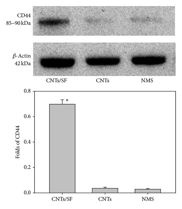
Figure 6.

Immunoreactive bands of CD44 and β-actin from fibroblast cells cultured on surfaces of CNTs/SF, CNTs, and NMS (nonmodified surface). The quantitative analysis of Western blotting was carried out using the ImageQuant-TL-7.0 software. These values that refer to the expression of CD44 were normalized by the expression of beta-actin.
