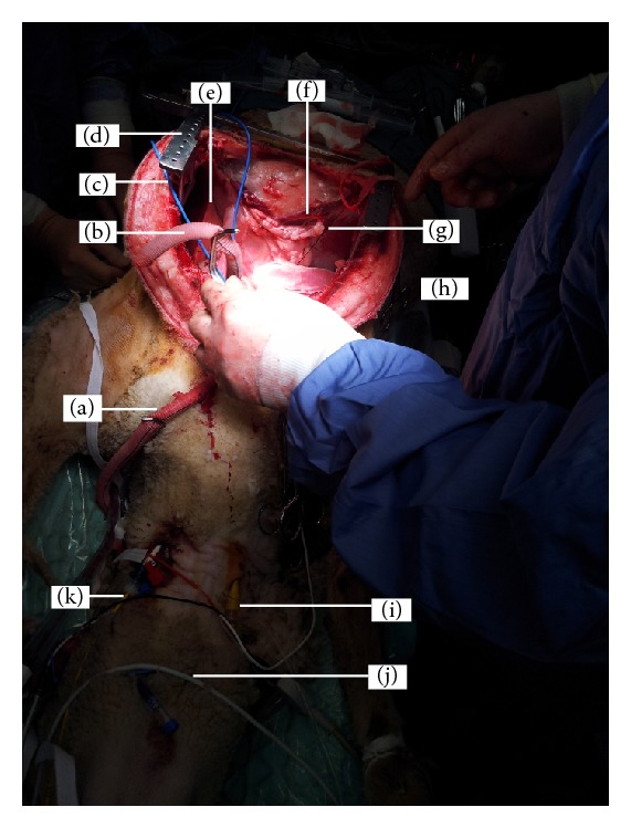
Figure 5.

Implanting an artificial heart in an ovine model. This study evaluated a biventricular mechanical device called BiVACOR that is being developed for patients with end stage heart failure. Sheep are anaesthetised and maintained as in humans undergoing cardiac transplantation. In this sheep model, anaesthesia is maintained by constant infusion of propofol and fentanyl. The approach to the heart is via a median sternotomy. Anastomoses to prosthetic blood vessels are created on the pulmonary vein (a) and artery (b) and dorsal aorta and clamped for subsequent attachment to the BiVACOR system (not pictured). In our completed trials, we attached grafts via an end-to-end anastomosis to the ascending aorta (16–18 mm) and pulmonary artery (20 mm) for left and right pump outflows, respectively. Inflow connections were achieved by removing the ventricles just below the atrioventricular groove (leaving about 1 cm of ventricular tissue) and suturing 38 mm grafts (end-to-end) to the ventricular tissue. All grafts were then attached to the pump. Blood flow after the prosthetic heart implant can be monitored by flow meters via probes (c). The surgical exposure of the chest cavity is maintained by a retractor (d) to visualise all organs. The right lung (e), diaphragm, and heart (h) are visible. A stay suture (g) facilitates the suspension of major blood vessels during dissection. Intravenous access for fluid administration was facilitated via a large bore catheter (i). Note the white colour of propofol (j) being used to maintain anaesthesia. During dissection and prior to the removal of the ventricles, continuous haemodynamic monitoring is achieved via the Swan-Ganz pulmonary artery catheter (k).
