
Figure 2.
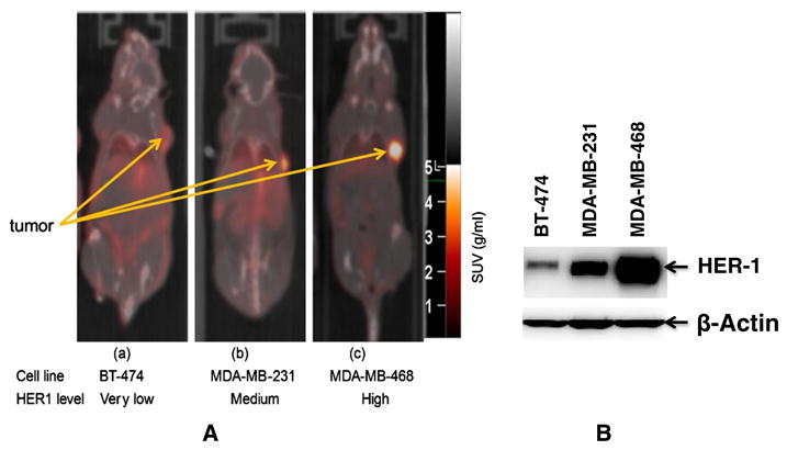
(A) Tumor uptake of 89Zr-panitumumab in various subcutaneous athymic nude female xenograft models. 10.18 ± 1.24 MBq of 89Zr-panitumumab were administered intravenously via tail-vein, and a 5-min CT scan followed by a 30-min static PET scan were performed at 96-h post-injection; (B) Comparative epidermal growth factor receptor (EGFR)-expression level of BT-474, MDA-MB-231, and MDA-MB-468 cell lines by western blot analysis. Cartesian molecular imaging software v5.0.2.30 and data are presented as EGFR/Actin ratio. The images are reprinted with the permission from Elsevier Inc. Bhattacharyya et al., Nucl. Med. Biol. 2013, 40, 451–457.