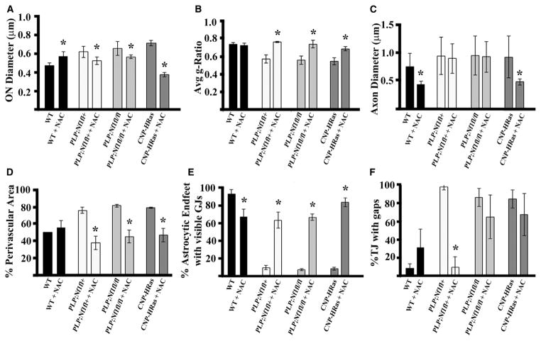
Figure 7. The Antioxidant NAC Rescues Myelin and Vascular Phenotypes in PLP;Nf1floxed and CNP-HRas Mice.
Quantification of phenotypes in WT, PLP;Nf1fl+, PLP;Nf1fl/fl, and CNP-HRas animals after 6 weeks of vehicle or NAC treatment, all in optic nerve 1 mm rostral to the chiasm.
(A) Optic-nerve diameter quantified from semithin cross-sections.
(B and C) Quantification of g-ratio (B) and axon diameter (C) of >1,000 axons/genotype measured by electron microscopy.
(D) Quantification of % perivascular area normalized to blood vessel area.
(E) Percentage of astrocyte endfeet with visible GJs (~300 GJs/animal).
(F) Percentage of endothelial TJs with gaps (~50–60 TJs/animal); n = 3–5 animals/genotype.

