Figure 1.
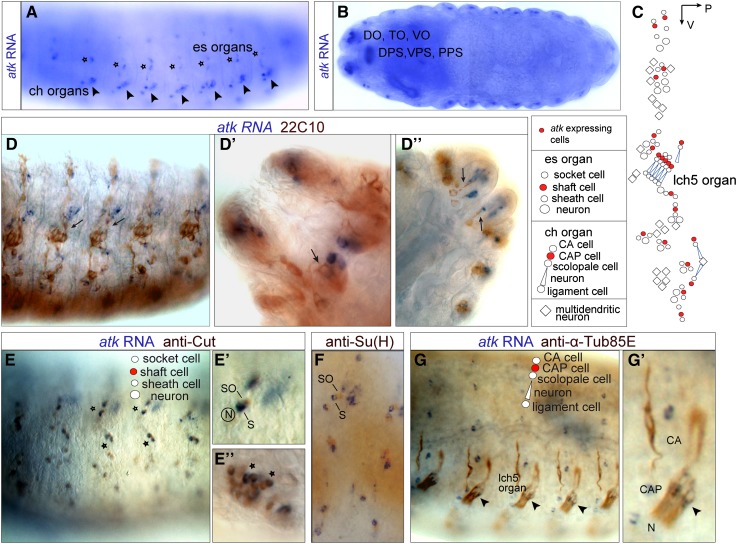
atk is expressed by an accessory cell in every ciliated sensory organ. (A and B) atk in situ hybridization in wild-type stage 16 embryos. (A) Lateral view showing atk in situ signal in a pattern consistent with expression in ch organs (arrowheads) and es organs (stars). (B) Horizontal view showing atk expression in chemosensory organs: dorsal organ (DO), terminal organ (TO), ventral organ (VO), dorsal, ventral and posterior pharyngeal organs (DPS, VPS, and PPS, respectively). (C) Schematic diagram of mechanosensory organs per hemisegment showing atk-expressing cells (red). (D–G) Embryos double-stained for atk in situ (blue signal) and different primary antibodies (brown signal). (D) atk is not expressed by ciliated neurons labeled by the neuronal marker mAb 22C10 (arrows, D–D′′). (E and F) In es and chemosensory organs, atk is expressed by the shaft cell: it colocalizes with anti-Cut signal (stars, E–E′′), which labels socket (SO) and shaft (S) cells, but does not colocalize with anti-Su(H), which labels the socket (F). (G) In ch organs, atk is expressed by the cap cell (CAP) based on atk-expressing cell position and colocalization with anti-α-Tub85E (arrowheads, G and G′). CA: cap attachment cell; N: neuron.

