Figure 3.
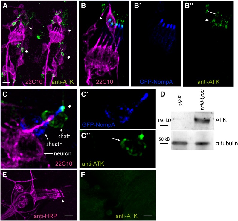
ATK localizes to the distal region of the dendritic cap in late embryonic mechanosensory organs. (A) Stage 16 embryonic hemisegments immunostained with the neuronal marker mAb22C10 (magenta) and with anti-ATK (green). ATK localizes apically to the sensory cilia in ch (arrowhead) and es (star) organs. (B and C) Detailed view of embryonic mechanosensory organs additionally showing GFP-NOMPA (blue) in the dendritic cap. Single channel images are shown for GFP-NOMPA (B′ and C′) and anti-ATK (B″ and C″). In lch5 (B) and es (C) organs, ATK localizes to the distal region of the dendritic cap (arrowhead in B, star in C), where it partially overlaps with GFP-NOMPA. The cytoplasm of cap and shaft cells are also stained (arrows). (D) Western blot with anti-ATK antibody. (E and F) ATK protein is not present in larval mechanosensory organs. Second instar larval lch5 organ stained with anti-HRP (E) shows no detectable anti-ATK staining. (F) Bars, 10 µm.

