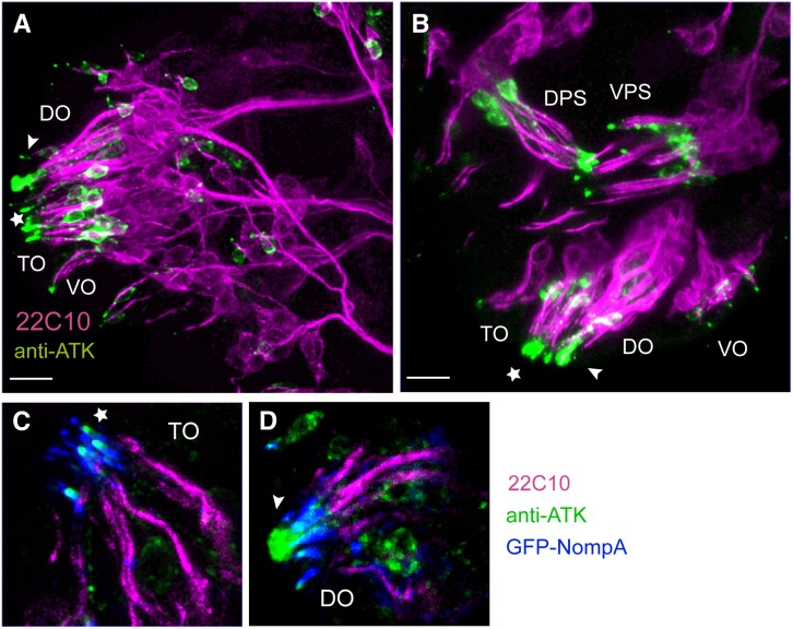
Figure 4.
ATK localizes to a supporting ECM in late embryonic chemosensory organs. (A and B) Stage 16 embryonic chemosensory organs immunostained with the neuronal marker mAb22C10 (magenta) and anti-ATK (green). DO: dorsal organ; TO: terminal organ; VO: ventral organ; DPS: dorsal pharyngeal organ; VPS: ventral pharyngeal organ. In DO (arrowhead) and TO (star), ATK localizes to the tip of the chemosensory cilia. Cell bodies of atk-expressing cells are also stained. Bars, 10 µm. (C and D) Detailed view of TO (C) and DO (D) additionally showing GFP-NOMPA (blue). (C) In TO, ATK localizes at the tip of the GFP-NOMPA region (star), where gustatory neurons are exposed to the environment. (D) In DO, ATK forms a vacuole-like structure in the center of the cilia cluster (arrowhead).

