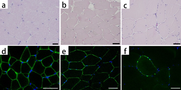
Fig. 2.

Hematoxylin and eosin staining (HE) and immunofluorescence staining for dystrophin: healthy pig (a, d), necrotic muscle (b, e) and the present pig (c, f). Dystrophin is stained light green. The nuclei are stained blue. The healthy pig shows small fibers (a), because of its young age and clear stainability at the sarcolemma (d). The necrotic muscle, which consists of muscle fibers that are massively necrotized because of an infarct, shows strongly eosinophilic pale cytoplasm and declined muscle nucleus stainability in HE (b), whereas dystrophin is stained at the sarcolemma (e). In the present pig, dystrophin is faintly and partially stained at the sarcolemma (f). Bar scales: 20 µm (a) and 50 µm (b–f).
