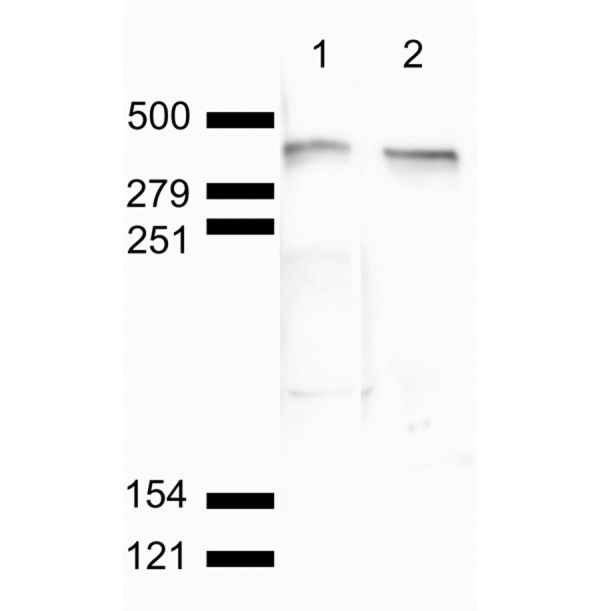
Fig. 3.

Western blot analysis of normal porcine muscle. Lane 1 is stained using pan-dystrophin antibody Dys1 (clone Dy4/6D3). Lane 2 is stained using the primary antibody (clone 1808) used in the present immunofluorescent examination. Closely spaced doublet bands of approximately 425 kDa were visualized on both lanes below the 500-kDa marker. Non-specific reactivity was not detected.
