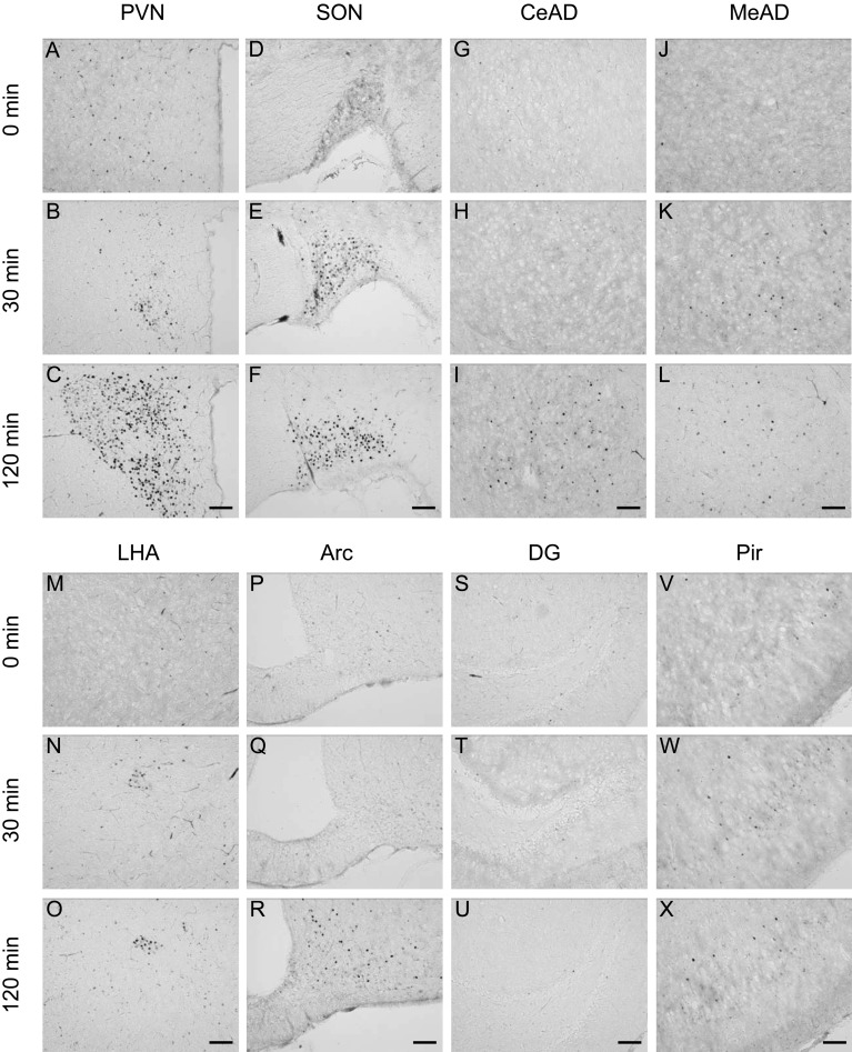
Fig. 2.
Immunohistochemical staining for c-Fos protein in the brain 0, 30 and 120 min after 2DG injection (400 mg/kg BW, ip). Representative photomicrographs of c-Fos-immunoreactive cells in the paraventricular nucleus (PVN, A–C), supraoptic nucleus (SON, D–F), central amygdaloid nucleus (CeAD, G–I), medial amygdaloid nucleus (MeAD, J–L), lateral hypothalamic area (LHA, M–O), arcuate nucleus (Arc, P–R), dentate gyrus (DG, S–U) and piriform cortex (Pir, V–X). Scale bars: 100 µm.

