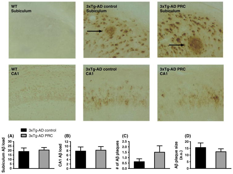
Fig. 4.
PRC regimen does not slow down Aβ accumulation in 3xTg-AD mice hippocampus. Representative images showing Aβ immunoreactivity in subiculum or CA1 hippocampus regions of 12.5–13.5-month-old WT control, 3xTg-AD control and 3xTg-AD PRC mice are shown. Aβ plaques are indicated by arrows. Quantification of Aβ accumulation by load values in subiculum and hippocampus CA1 regions is showed in (A) and (B), respectively. Number and size of Aβ plaques are shown in (C) and (D). [10–12 (A, B, C) and 5–7 (D) samples per group].

