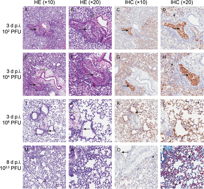
Figure 2.
Histopathologic changes in lungs of mice infected with human influenza A/Anhui/1/2013 (H7N9) virus. Mice (n = 3, each day postinfection [p.i.]) were infected with 102–106 plaque-forming units (PFU)/mouse of A/Anhui/1/2013 (H7N9) virus and killed on day 3 (A–L) or day 8 p.i. (M–P). Mouse lungs were fixed in 10% neutral buffered formalin and stained with hematoxylin-eosin (HE) (A, B, E, F, I, J, M, and N) or immunohistochemical staining (IHC) with anti–influenza A nucleoprotein antibody (C, D, G, H, K, L, and O), or Masson trichrome staining was performed to visualize hyaline membrane formation (P). Magnification ×10 (A, C, E, G, I, K, M, and O), ×20 (B, D, F, H, J, L, N, and P). A, B, E, F, I, and J, Necrosis of bronchiole with cell debris in bronchiolar lumen (solid arrow). Alveolar collapse and enlargement of alveolar ducts. Alveoli containing edema (open arrow) and inflammatory cells. C, D, G, H, K, and L, Solid arrow indicates influenza antigen-positive cell debris in bronchiolar lumen and influenza antigen-positive staining in respiratory epithelial cells (a). O, Weak influenza virus antigen-positive staining in bronchiolar (solid arrow) and in respiratory epithelial cells (a), including collapsed areas. P, Masson trichrome stain showing hyaline membrane formation lining alveolar ducts (solid arrow) and alveolar collapse (open arrow).

