
Figure 2. Nanostructure and supramolecular cohesion within PA 1 and PA 2 assemblies.
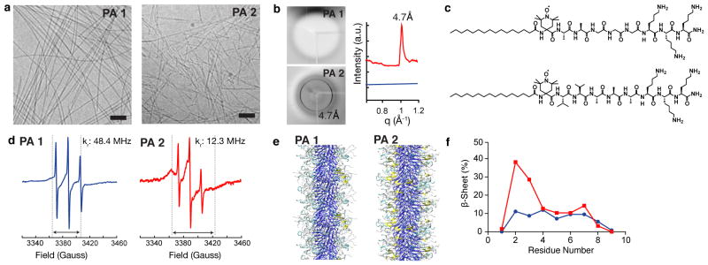
(a) Cryogenic TEM showing representative nanostructures in cell media. Scalebars: 100 nm. (b) X-ray diffraction of solutions of PA 1 (blue) and PA 2 (red). 4.7 Å corresponds to β-sheet hydrogen bonding. (c) Chemical structures of the spin labeled analogues of PA 1 (top) and PA 2 (bottom) with a site specific spin label (TOAC) located at the first amino acid adjacent to the fatty acid tail to probe the β-sheet hydrogen bonding segment of the assemblies. (d) Electron paramagnetic resonance spectra of PA 1 and PA 2 combined with 20% of the corresponding spin labeled analogue. Dashed lines and double headed arrows indicate the spectral broadening that and PA 2 occurs with reduced rotational diffusion (kr). (e) Atomistic modeling of PA 1 demonstrating differences in β-sheet hydrogen bonding (yellow: β-sheet hydrogen bonding, cyan: β-turn, gray: random coil, blue: alkyl tail). (f) Data from modeling experiments to determine the distribution of β-sheet hydrogen bonding within assemblies of PA 1 (blue) and PA 2 (red) as a function of residue number from the alkyl tail.