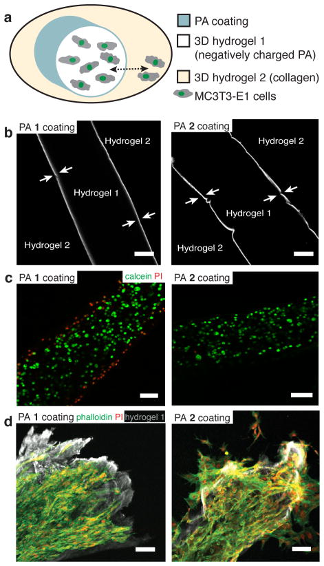
Figure 5. Creating a cell barrier with PA coatings to compartmentalize cells.
(a) A schematic of the experimental setup. A cell-encapsulated hydrogel (palmitoyl-VVAAEE 1%, w/v, hydrogel 1) is surrounded by collagen (hydrogel 2) with PA 1 or PA 2 coating at the interface. The experiment evaluates the cell migration through the hydrogel interface from hydrogel 1 to hydrogel 2 (dashed double headed arrow). (b) A representative confocal slice from the two-compartment hydrogel system (as shown in (a), but without cells) where hydrogel 1 is coated with either fluorescently labeled PA 1 or fluorescently labeled PA 2 (arrows indicate the coating layer which is approximately 10 μm thick). Scalebars: 100 μm. (c) Viability of MC3T3-E1 cells that were encapsulated in the hydrogel 1, coated with either PA 1 or PA 2, and stained after 1 hour with calcein (green, live cells) and propidium iodide (red, dead cells). Scalebars: 100 μm (d) PA 1 or PA 2 coated cell encapsulating hydrogel 1 were further embedded in hydrogel 2 (as in the illustration in (a)) and cultured for 7 days, before staining for actin (phalloidin, green) and nuclei (propidium iodide, red). Cells remained confined within the hydrogel 1 compartment (grey) when coated with PA 1, while cells escaped into the surrounding collagen when coating was done with PA 2. Scalebars: 100 μm.

