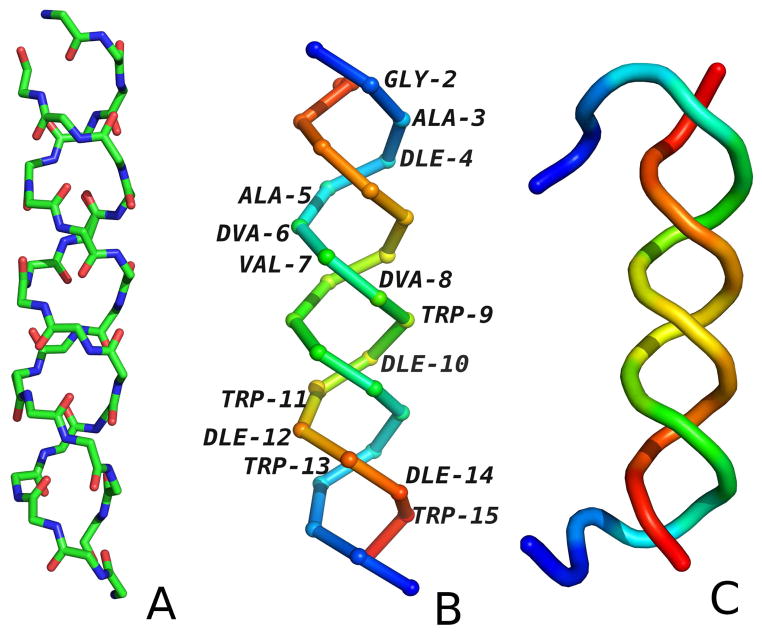
Fig. 8.
The experimental structure of gramicidin D (PDB code: 1AL4). (A) The backbone with all atoms shown to indicate the hydrogen-bonding contacts, (B) The Cα trace with residues colored from blue to red from the N-terminus to the C-terminus for each chain. (C) The cartoon representation of structure obtained from MREMD simulation at 250K colored from blue to red from the N-terminus to the C-terminus for each chain.

