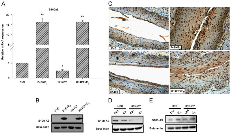
Fig. 5.
The 17β-estradiol enhances the expression of S100-A9. Quantitative real-time PCR analysis showed a higher mRNA expression of S100a9 inK14E7+E2 and FvB +E2 mice compared to untreated mice (FvB or K14E7) (A). Bars (relative expression) represent the mean±SD of three independent experiments (*p<0.05, **p<0.01, Student's t-test) normalized to Hprt mRNA and compared with the control (untreated FvB mice). Western blot analysis (B) and immunohistochemistry (C) also revealed a marked up-regulated S100-A9 expression in K14E7+E2 and FvB +E2 mice. S100-A9 positive cells in immunohistochemistry show brown staining in the nucleus and cytoplasm. The results show a representative of three experiments, nuclei were counterstained with hematoxylin and visual field at 40 × magnification. Western blot analysis of Human primary and HPV16 E7 transformed keratinocytes treated with vehicle or ICI 182,780 (50 nM for 48 h) (D) and vehicle or E2 (10 nM) (E) for 24 h after 48 h starvation in the absence of any growth factor. Cell fractionation was conducted, and lysates were subjected to SDS-PAGE followed by Western blot with anti-S100-A9 and anti-beta-actin (control) antibodies. All gene and protein symbols were written according to the HUGO Gene Nomenclature Committee (HGNC) and UniProtKB, respectively.

