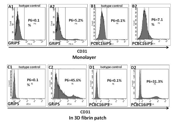
Figure 2. The efficiency of hiPSC-EC differentiation increases when the cells are encapsulated in a fibrin patch.
hiPSCs were differentiated (A1-B2) in monolayers or (C1-D2) after suspension in a fibrin patch. Differentiation efficiency was evaluated by comparing (A2, B2, C2, D2) CD31 expression with isotype controls (A1, B1, C1, D1) via flow cytometry. The maximum efficiencies obtained with each differentiation protocol are shown for both GRiPS- and PCBC16iPS-lineage cells.

