
Figure 6.
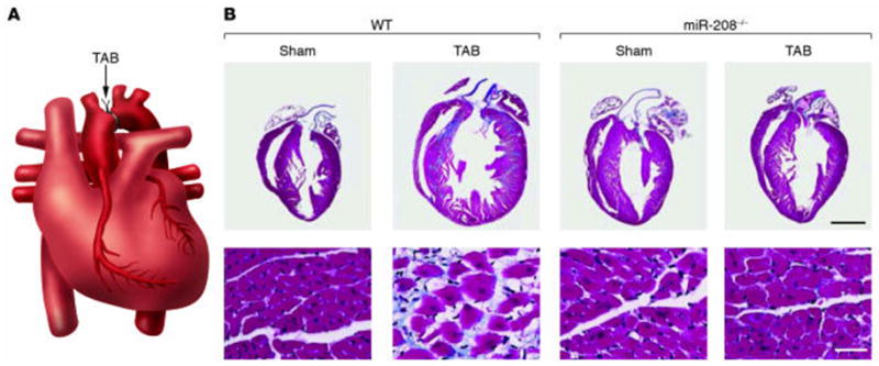
Requirement of miR-208 for cardiomyocyte hypertrophy and fibrosis. (A) Schematic diagram of a heart following thoracic aortic banding (TAB). (B) Sections of hearts of approximately 3-month-old wild-type and miR-208−/− mice are shown following sham operation or TAB for 21 days. High-magnification views of the ventricular wall are shown at the bottom. Trichrome staining identifies fibrosis in blue. Note that hypertrophy and fibrosis are diminished in miR208−/− mice compared with wild-type following TAB. (Reproduced with permission from van Rooij and Olson, J. Clin. Invest. 2007; 117:2369–2376).