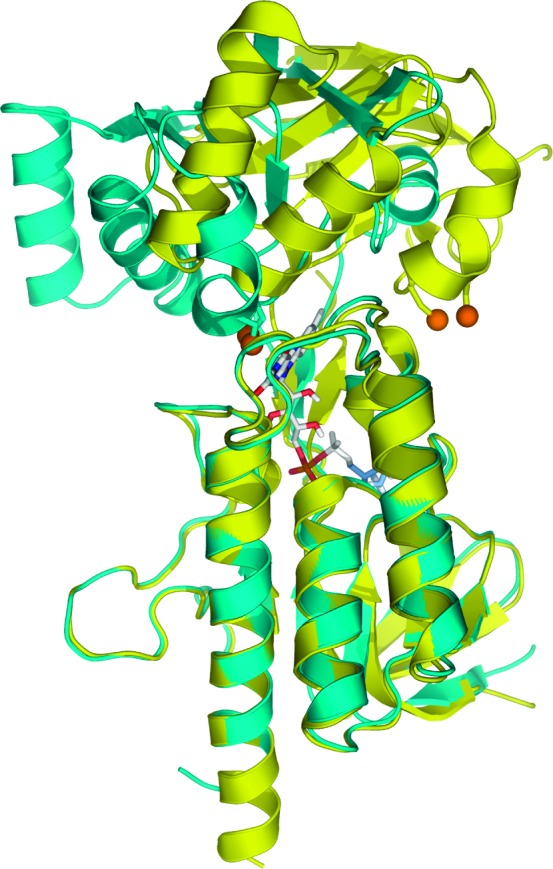
Figure 1.

The FO and FR conformations of ecTrxR, superimposed in cyan and yellow, respectively. Orange spheres are used to represent the disulfide-forming cysteines in the FO conformation, and the cysteine/serine pair is used in the crystallographic trapping of the ecTrxR FR conformation. The image was constructed from the FO and FR PDB files 1TDF and 1F6M, respectively.
