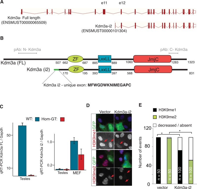
FIGURE 4:
Kdm3a encodes two protein isoforms. (A) Intron-exon diagram of Kdm3a. Short isoform i2 was cloned following ENSEMBL prediction (ID numbers between parentheses). First and unique Kdm3a-i2 exon is contained within intron 11. (B) Diagram illustrates the amino acid position of Kdm3a protein domains in full-length (FL) and isoform 2 (i2). Green line, i2-exclusive exon; amino acid sequence shown in bold. pAb indicates the relative position of amino terminal Kdm3a single polyclonal antibody used along this study. (C) qRT-PCR from testis and MEF RNA show a decrease of Kdm3a-FL and unaltered transcript levels of i2 in Kdm3aGT/GT testis. A slight reduction of Kdm3a-i2 is observed in Kdm3aGT/GT primary MEFs. (D) RPE1 cells transfected with GFP tag fused upstream of Kdm3a-i2. Monoclonal antibodies to mono- or dimethylated Lys-9 of histone H3 were used to determine the state of histone methylation (red) in transfected cells (green). Arrows point to transfected cells. (E) Quantitation of (D). n = number of cells counted for each condition. *p < 0.05, Fisher's exact test.

