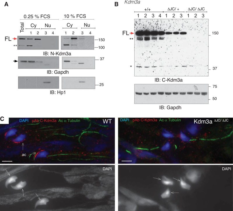
FIGURE 7:
Cytoplasmic distribution of endogenous Kdm3a. (A) Subcellular fractionation of serum-starved and exponentially growing MEFs immunoblotted with the indicated antibodies. Sequential disruption of cell and nuclear membranes gives rise to two cytoplasmic fractions: soluble (1) and membrane (2) and two nuclear fractions: soluble (3) and cytoskeletal (4). GAPDH and HP1 are controls for cytoplasmic and nuclear fractions, respectively. Red and black arrows indicate FL-Kdm3a and GAPDH proteins, respectively. (B) Total extracts from four wild-type (WT) and 6 ΔJC Kdm3a mice immunoblotted with pAb antibody directed to the C-terminal region of Kdm3a (see also Figures 6B and S2A). The immunizing peptide of this commercially available antibody is likely contained within ΔJC deletion, as no band is observed in any of the three Kdm3aΔJC/ΔJC mice analyzed. GAPDH is used as loading control. ** and * indicate potentially nonspecific bands. (C) Immunohistochemistry shows Kdm3a localization in acrosomes (ac) of wild-type (WT) mature spermatids but not Kdm3aΔJC/ΔJC as detected with C-Kdm3a antibody described in (B). Arrows indicate the dorsal surface of the spermatid on DAPI-stained nuclei.

