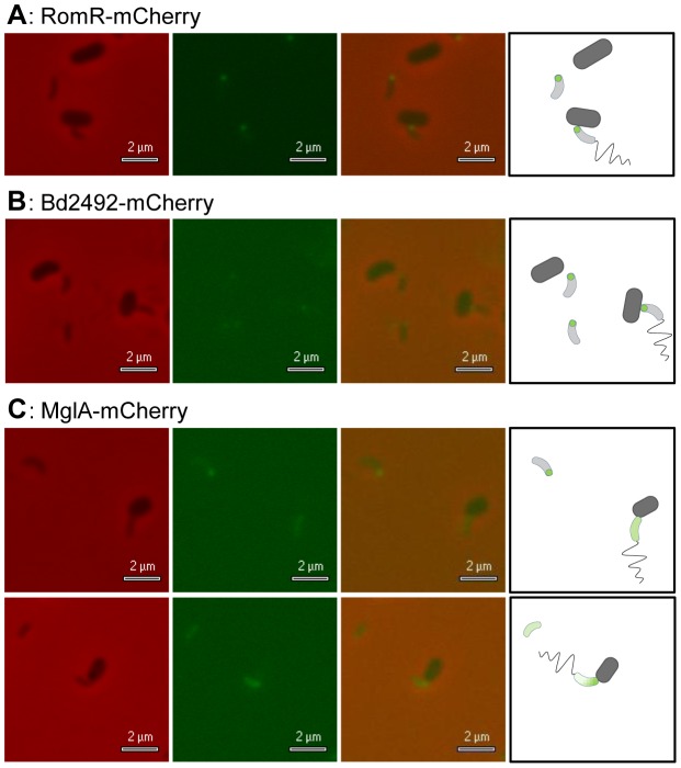
Figure 6. B. bacteriovorus RomR-mCherry and Bd2492-mCherry localised at the prey-interaction pole; MglA-mCherry showed variable diffuse foci.
B. bacteriovorus cells were incubated with E. coli S17-1 prey cells for 5 minutes, allowing sufficient time for some of the Bdellovibrio cells to attach to prey. Panels- A: The lower prey-cell shows a typical attached Bdellovibrio cell, with a RomR-mCherry focus at the anterior (attached) pole of the Bdellovibrio. B: The rightmost prey-cell shows a typical attached Bdellovibrio cell, with a Bd2492-mCherry focus at the anterior (attached) pole of the Bdellovibrio. C: MglA-mCherry Bdellovibrio cells had variable foci, including diffuse and unipolar localisations. From left to right, all panels show brightfield, fluorescent, and merged images and a graphical representation. Fluorescent exposure = 2 seconds.

