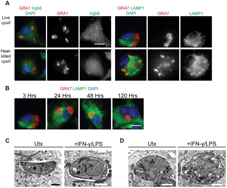
Figure 2. The fate of heat-killed and live cpsII parasites in host cells.
C57BL/6 bone marrow derived macrophages were infected with cpsII parasites and examined using immunofluorescence assays (a). Bone marrow-derived macrophages activated with IFN-γ (100 U/ml) and LPS (0.1 ng/ml) were infected with freshly lysed or heat-killed parasites for 3 hours. Intracellular parasites were stained for host Irgb6 or LAMP1 recruitment in green. Parasites were stained with a mouse monoclonal antibody to GRA1 to identify the parasitophorous vacuole or rabbit polyclonal sera against GRA7 in red. IFN-γ and LPS activated bone marrow-derived macrophages were infected with freshly lysed cpsII parasites and fixed at 3, 24, 48 and 120 hours post-infection (b). Parasite vacuoles were identified with rabbit polyclonal sera to GRA7 (red) and host LAMP1 was identified with a rat monoclonal antibody. Scale bar = 10 µm. Electron micrograph images of infected macrophages treated with IFN-γ (50 units/ml) and LPS (10 ng/ml) or untreated at 2 hours post-infection (c). Parasites persist in intact vacuoles and do not display blebbing or disruption of the parasitophorous vacuole. Some cpsII parasites were found to exhibit non-productive cell division in IFN-γ and LPS- treated or untreated macrophages when examined 24 hours post-infection (d). Scale bars = 1.5 µm.

