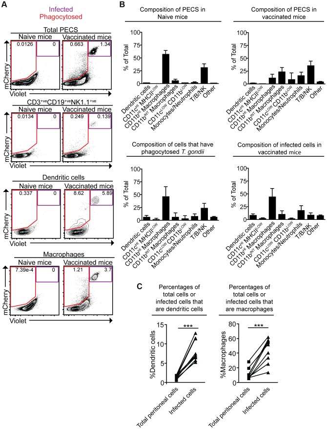
Figure 3. Composition of total cell populations, mCherry+veViolet−ve cell populations, and mCherry+veViolet+ve populations from the PECS of naïve and vaccinated mice.
Mice were vaccinated with 106 Violet-labeled, mCherry-expressing cpsII parasites intraperitoneally and sacrificed 18 hours post-vaccination. Cell type composition of total peritoneal cell populations in naïve and vaccinated mice, and the cell type composition of mCherry+veViolet−ve cells and mCherry+veViolet+ve cells in vaccinated mice were examined. Representative flow plots demonstrating infected cells and cells that have phagocytosed T. gondii for each major cell type present in the PECS are shown (a). The composition of the PECS in naïve mice and vaccinated mice, and the composition of infected cells (mCherry+veViolet+ve) and cells that have phagocytosed T. gondii (mCherry+veViolet−ve) are depicted (b). Percentages of macrophages and dendritic cells in the total peritoneal cell population in vaccinated mice are compared to the percentages of infected cells that are macrophages and dendritic cells (c). T/B/NK cells are identified by expression of CD3, CD19, or NK1.1. Dendritic cells were identified as CD3−ve,CD19−ve,NK1.1−ve,CD11cHI,MHCIIHI. Monocytes and neutrophils were defined as CD3−ve,CD19−ve,NK1.1−ve,CD11cLOW-INT,Gr-1+ve. Macrophages were identified as CD3−ve,CD19−ve,NK1.1−ve,CD11cLOW-INT,Gr-1−ve,CD11bINTorHI. *p<0.05; ***p<0.0005. AVG±STDEV. A paired, two-tailed student's t test was used to analyze the data in (c). Results shown are from one representative experiment. Similar results were obtained over the course of seven separate experiments.

