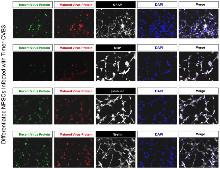
Figure 6. Inspection of recent and matured virus protein expression in differentiated NPSCs infected with Timer-CVB3.
NPSCs cultures were differentiated for five days, and then infected with Timer-CVB3 for three days, followed by immunostaining for lineage markers (white signal). “Fluorescent timer” protein produced early on (matured virus protein) marked cells which were infected early on. Newly infected cells were distinguished by green fluorescence (recent viral protein).

