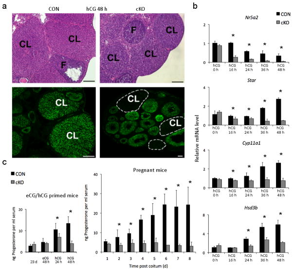
Figure 1. Deletion of Lrh-1 in the peri-ovulatory follicle via PrCre causes luteal insufficiency.
(a) Corpora lutea (CL) and follicles (F) in ovaries from CON (Nr5a2fl/fl) and cKO (Nr5a2fl/fl;PrCre/+) mice at 96 h after treatment with equine chorionic gonadotropin (eCG) to induce follicle development and 48 h after treatment with 5 IU human chorionic gonadotropin (hCG) to induce ovulation. Lower panels demonstrate ablation of the Lrh-1 signal in corpora lutea and its retention in follicles. Scale bars, 200 μm. (b) Abundance for Lrh-1 (Nr5a2) mRNA and the steroidogenic proteins Cyp11a1, Star and Hsd3b, determined by qPCR in granulosa cells aspirated prior to induction of ovulation with hCG (designated hCG 0h) and laser microdissected corpora lutea from CON and cKO mice at 0–48 h after hCG (n=3–10 mice per point) (c) Left panel: serum progesterone in immature CON and cKO mice prior to (23d), 48 h after gonadotropin induction of follicle development and 24 and 48 h after hCG. Right panel: peripheral progesterone concentrations in CON and cKO mice for the first eight days post copulation (dpc). Values are means ± SEM, n=5–10 mice, asterisks designate differences between cKO and CON animals at each time point (p<0.05).

