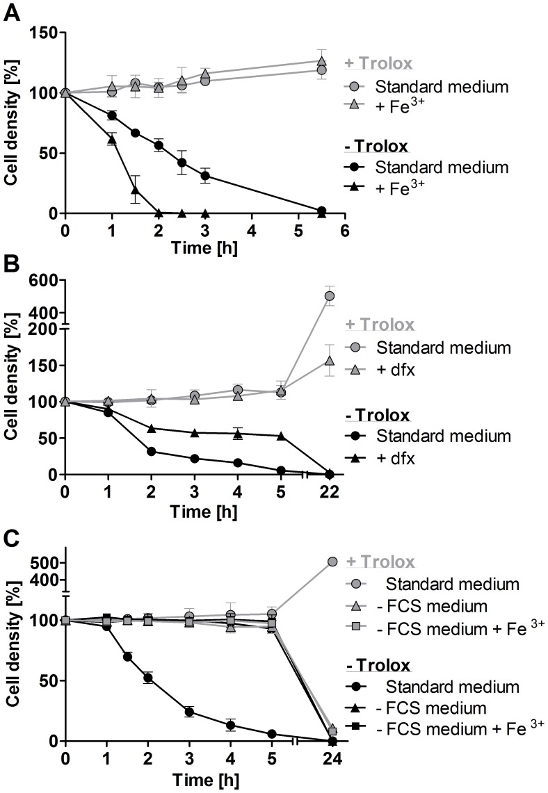
Figure 3. Exogenous iron promotes lysis of the px I–II −/− BS cells.
A. The mutant parasites were incubated for up to 5.5°C in standard medium ±100 µM Trolox in the presence and absence of 100 µM Fe3+. WT cells behaved like the mutant cells in the presence of Trolox. B. The px I–II−/− cells were incubated for 5 h at 22°C and then cultured overnight at 37°C in medium ±100 µM Trolox in the presence and absence of 100 µM deferoxamine (dfx). WT behaved like the mutant cells in the presence of Trolox. C. The px I–II−/− cells were incubated for 5 h at 19°C and then cultured overnight at 37°C in medium ±10% FCS and/or 100 µM Trolox in the presence and absence of 100 µM Fe3+. The behavior of WT cells in the absence of FCS was identical to that of the mutant cells. At the different time points, living cells were counted. The values represent the mean ± SD of three independent experiments.

