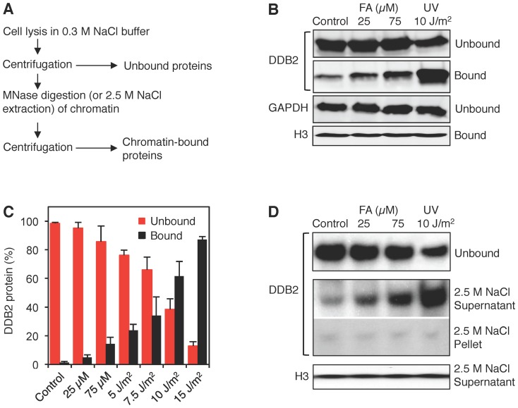
Figure 3. Association of UV-DDB with damaged chromatin.
(A) Flow diagram illustrating how chromatin was dissected to monitor the binding of UV-DDB. Unbound proteins were released by salt (0.3 M NaCl) extraction and the remaining chromatin was solubilized by MNase digestion. (B) Western blot visualization of the chromatin partitioning of UV-DDB using antibodies against DDB2. GAPDH (glyceraldehyde 3-phosphate dehydrogenase), marker of unbound proteins; histone H3, marker of chromatin. Human fibroblasts were exposed for 18 h to formaldehyde or UV-irradiated at the indicated doses. (C) Quantification of three independent binding assays demonstrating the differential interaction of DDB2 with formaldehyde- and UV-damaged chromatin (error bars, S.D.). (D) Release of chromatin-bound DDB2 and histone H3 by high-salt extraction. After incubation with 0.3 M NaCl buffer, the chromatin was dissolved with 2.5 M NaCl, thus liberating non-covalently bound chromatin proteins.

