
Figure 6.
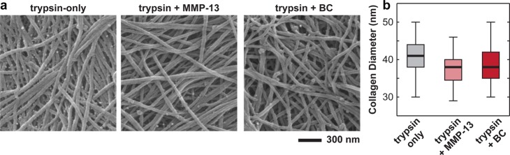
(a) Scanning electron microscopy images of trypsin-treated cartilage disk surfaces, prepared via Ohtani’s procedure25 to retain its 3D architecture, including trypsin only, trypsin + MMP-13, and trypsin + bacterial collagenase treated disks. (b) Box-and-whisker plot of the distribution of collagen fibril diameters measured for the three types of disks (n ≥ 100 fibrils for each treatment).