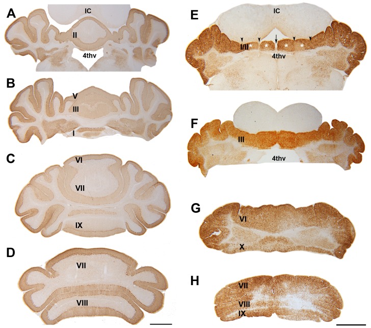
Figure 3. Frontal sections of P21 wild type (A–D) and nax (E–H) cerebella immunostained with CaBP.
A–D) The wild type cerebellum shows normal lobules and the central vermis is prominent with hemispheres on either side. E–H) From the rostral section (E) to the caudal section (H), the nax cerebellum shows obvious signs of a hypoplastic vermis compared to the wild type. In lobules I/II (E), the hemispheres fail to fuse completely in the midline, but fusion becomes more obvious as the sections progress posteriorly. The underdeveloped anterior lobules of the nax cerebellum have shallow rostrocaudal fissures (arrow head) causing a blocked appearance of the cortex (white asterisk) that is symmetrical about the midline (arrow). A putative vermis area may be present in caudal sections of the nax cerebellum, but all that is noticeable is that the medial part (putative vermis) is smaller in relation to the hemispheric part, compared to the wild type. Individual lobules in the vermis are indicated in Roman numerals. Abbreviation: 4thv = fourth ventricle; IC = Inferior colliculus. Scale bar: D = 1 mm (A–D); H = 1 mm (E–H).

