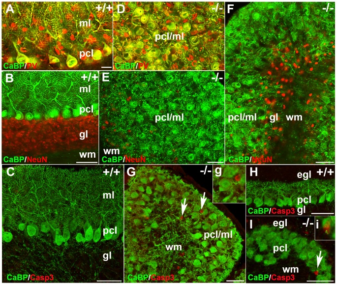
Figure 4. Transverse sections at P21 and sagittal sections at P6 and P17 of the nax mutant and wild type cerebellum.
A) Double immunostaining with CaBP (green) and PV (red) in a transverse section through the cerebellum of the wild type sibling showing inhibitory interneurons and the dendrites of Pcs in the molecular layer (ml) and Purkinje cell soma in the Purkinje cell layer (pcl). B) A transverse section through the cerebellum of the wild type sibling double immunostained with CaBP (green) and NeuN (red), showing the three layers of the cortex: molecular layer (ml), Purkinje cell layer (pcl) and granular layer (gl). C) Double immunostaining with CaBP (green) and cleaved caspase 3 (red) in a sagittal section of the wild type sibling cerebellum at P17 showing lack of caspase + cells in cerebellum. D) Double immunostaining with CaBP (green) and PV (red) in a transverse section through the cerebellum of the nax mutant showing that the Pcs fail to form a uniform monolayer. Inhibitory interneurons and Pcs soma are intermingled in the ml; labeled as pcl/ml. E–F) Transverse sections through the cerebellum of the nax mutant, anteromedially (E; putative anterior vermis) and posterolaterally (F; hemisphere). In the anterior lobe, an apparent lack of the granular layer places the pcl/ml directly in contact with white matter (D). In the posterolateral cerebellum, a small amount of cerebellar granule cells form a granular layer between the pcl/ml and wm (E). Purkinje cells are arranged in different directions. G) Double immunostaining with CaBP (green) and cleaved caspase 3 (red) in a sagittal section through the cerebellum of the nax mutant at P17 showing that the caspase 3 immunopositive cells (arrow; higher magnification in “g”) are not co-labeled with CaBP+ (Pcs) cells. H–I) Double immunostaining with CaBP (green) and cleaved caspase 3 (red) in sagittal sections of wild type cerebellum (H) and nax mutant cerebellum (I) at P6 showing that the caspase 3 immunopositive cells (arrow; higher magnification in “I”) are not co-labeled with CaBP+ (Pcs) cells. Scale bar: A = 25 µm (A, D); B, E, F = 50 µm, C, H, J = 50 µm, G = 40 µm.

