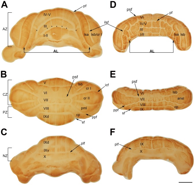
Figure 5. Whole mount immunostaining of wild type (A–C) and nax (E–F) cerebella at P8 with PLCβ4.
Individual lobules in the vermis are indicated in Roman numerals. A–C) The central vermis and cerebellar hemispheres are normally lobulated in the wild type cerebellum. A) The anterior lobe (AL; lobules I–V) is separated from the rest of the cerebellum by the primary fissure (pf). Wide PLCβ4 immunopositive stripes separated by narrow immunonegative stripes are clearly seen in the anterior vermis (indicated by arrowheads). B) The dorsal aspect of the cerebellum shows the posterior lobe (lobules VI–IX). The lobulus simplex (lsa and lsb) is separated from the ansiform lobules (cr I and cr II; crus I and crus II) by the posterior superior fissure (psf). Lobule VIII is separated from lobule VII by the prepyramidal fissure (ppf) and its lateral extension as the copula pyramidis (cp) is separated from lobule IX by the secondary fissure (sf). The vermis of lobule VI–VII is immunonegative with some extension of stripes from the rostral and caudal regions indicating this as a transitional zone. In lobule VIII and IXd, PLCβ4 is expressed in stripes. C) The ventral aspect shows the posterolateral fissure (plf) between lobule IX and X. PLCβ4 is expressed in stripes in lobule IXv and lobule X is immunonegative. D–F) D) The small, underdeveloped nax cerebellum shows severe anomalies in the anterior lobe that also extend across the primary fissure into lobule simplex “a” (lsa). PLCβ4 is expressed uniformly in the underdeveloped anterior lobe with no obvious stripes. E) From the dorsal aspect, the expression pattern of PLCβ4 shows the presence of a vermis. The vermis of lobule VI and VII is PLCβ4 immunonegative, whereas, their lateral extensions as the lobule simplex b and undivided ansiform lobule appear to have a striped expression pattern. Expression of PLCβ4 is in stripes in lobule VIII and dorsal lobule IX. Lobule X is PLCβ4 immunonegative. Abbreviations: ilsf = intralobule simplex fissure, icf = intercrural fissure. Scale bar: 1 mm.

