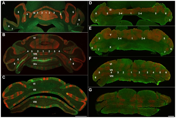
Figure 7. Number of stripes is compared in the wild type (A–C) and nax cerebellum (D–G) at P24.
Stripe markers are PLCβ4 (red) and ZII (green). A–C) Transverse sections through the anterior and posterior lobes of the wild type cerebellum show five clear ZII+ stripes in the vermis that alternate with PLCβ4+ stripes. The stripes in the hemispheres are not as clear as in the vermis, but they are distinguishable and numbered accordingly. D–G) In the nax mutant cerebellum, fewer stripes of typical gene expression were found. D) ZII+ stripes in the anterior lobe vermis are weak or absent, but are stronger in the hemispheres. E) The pattern of stripes is clearly present in the caudal anterior lobe of the nax mutant, but it appears that either stripes 2–4 are fused or a number of stripes are missing. F, G) The number of stripes in the caudal part of the posterior lobe appears to be similar to the normal pattern. The lobules are indicated by Roman numerals. The ZII+ Purkinje cell stripes are labeled (P1+ to P7+ as 1–7 for clarity). Scale bar: C = 1 mm (A–C); D, E, F, G = 1 mm.

