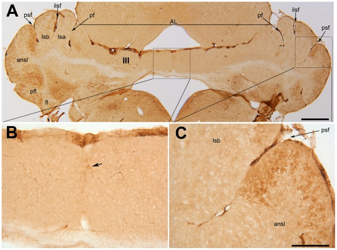
Figure 8. Transverse section through the anterior lobe of the nax cerebellum at P19 immunostained with ACP2.
A–C) A section through lobule III of the nax cerebellum shows the underdeveloped cerebellum, particularly in the vermis, and the distinguishable primary fissure (pf) and lobules in the lateral cerebellum (A). Acp2 is clearly expressed in stripes in the hemispheres, seen at low magnification in A and at high magnification in C. Although there are no stripes in the putative vermis at low magnification (A), higher magnification shows a few Purkinje cells in the midline that express Acp2 (arrow) (B). Abbreviations: AL = anterior lobe, ansl = ansiform lobule, fl = flocculus, ilsf = intralobule simplex fissure, icf = intercrural fissure, Ls = lobulus simplex, pfl = paraflocculus, psf = posterior superior fissure. Scale bars: A = 1 mm; C = 250 µm (B–C).

