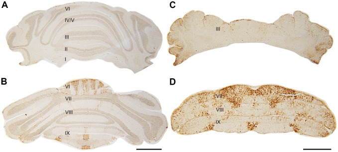
Figure 9. Transverse sections of wild type (A–B) and nax (C–D) cerebella at P19 immunostained with HSP25.
Individual lobules are indicated in Roman numerals. A–B) HSP25 immunostaining is absent in the AZ and PZ, and expressed in parasagittal stripes in the CZ and NZ of the wild type cerebellum. C–D) The nax cerebellum shows similar staining to the wild type in the AZ and PZ, however, uniform expression occurs in the CZ (lobule VII). Lobule IX is elongated, but has a similar pattern of HSP25+ stripes as the wild type. Scale bar: B = 1 mm (A–B); D = 500 µm (C–D).

