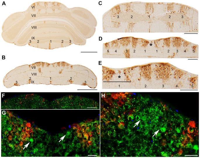
Figure 10. Transverse sections of wild type (A,C) and nax (B,D–E) cerebella at P22 immunostained with HSP25.
A,C) HSP25 immunostaining shows an array of parasagittal stripes in the CZ and NZ of the wild type cerebellum, with high magnification of the CZ shown in C. B, D– E) The nax cerebellum shows similar staining to the wild type in the elongated lobule IX. In the CZ several parasagittal stripes are present, however, about five stripes occur on each side of the midline (asterisk; D is a higher magnification of B). At higher magnification E, the midline stripe (asterisk) is clearly absent, which could be due to the lack of complete fusion across the midline (asterisk) or from a split occurring later on during development. F–H) Transverse section of the nax cerebellum at P21 immunolabeled with HSP25 (red) and CaBP (green) (higher magnification in G and H). In the nax mutant, CaBP+ Purkinje cells (arrows) and DAPI+ nuclei (blue) are located between HSP25+ stripes in the CZ. Scale bars: A = 1 mm; B = 1 mm; C = 250 µm; D = 250 µm; E = 250 µm; F = 250 µm; G and H = 40 µm.

