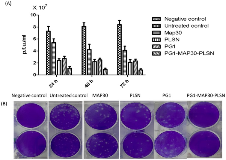
Figure 5. Evaluation of the peptides antiviral activities using plaque formation assay.
(A) Virus load expressed as plaque forming units per ml (p.f.u/ml) was significantly reduced after treatment with all the peptides compared to untreated cells. The peptide-fusion protein (PG1-MAP30-PLSN) showed the highest inhibition potential compared with the other peptides. (B) Plaque formation assay shows the reduction of plaque generation after the treatment of the infected cells with the peptides. (Two-way ANOVA with Bonferroni post-test, p<0.001).

