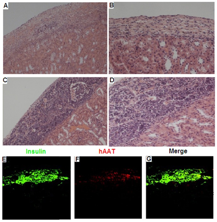
Figure 5. Pathological HE staining and immunostaining of the left kidneys after transplantation.
NIT-1 group at 14 d and 28 d after transplantation. Fourteen days after the transplantation, only a small number of transplanted NIT-1 cells remained in the kidneys. No transplanted NIT-1 cells were observed after 28 d, showing that the cells occurred fibrosis(A, B). NIT–hAAT group at 14 d and 28 d after transplantation, Fourteen days after transplantation, most of transplanted NIT–hAAT cells were still visible in renal capsule. After 28 days, there were no significant changes in the numbers of infiltrating inflammatory cells and a number of the transplanted NIT–hAAT cells were still present in the renal capsule(C,D). (scale bars, 200 µm). Immunostaining was performed at 28 d after transplantation NIT-hAAT cells under the kidney capsule. After rehydration, sections were incubated with anti-insulin and anti-hAAT antibodies. Insulin and hAAT at 28 d after transplantation are shown(E,F,G). (scale bars, 50 µm).

