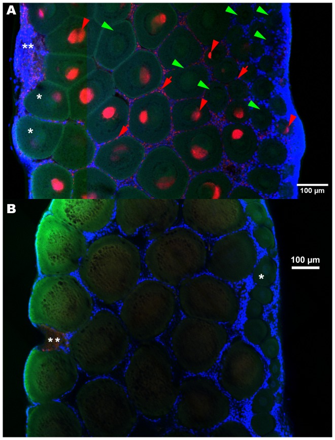
Figure 3. FISH detection of Wolbachia in ovaries, infected (A) versus control, uninfected (B).
The germaria are on the right borders, areas undergoing degradation on the maturing side of the ovaries (**). Oocyte nuclei are sometimes visible as dots (meiotic prophase) (*). A: The Wolbachia were present in oocytes of all sizes (red arrowheads) and their follicle cells (red arrows), though many oocytes remained uninfected (green arrowheads), especially the smaller ones. B: In uninfected ovaries. Red: Wolbachia FISH probe W1,2-Cy3, green: phalloidin, blue: DAPI.

