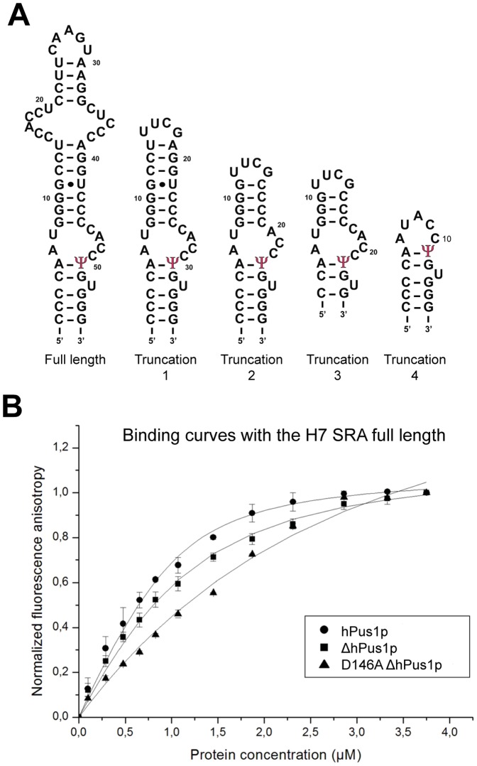
Figure 1. Human pseudouridine synthase 1 binds to various SRA constructs.
(A) Secondary structures and sequences of the SRA constructs used in the binding tests with the putative U to Ψ position indicated. (B) Fluorescence anisotropy measurement of SRA binding to different hPus1p constructs. Fluorescein-conjugated SRA sequences were incubated with increasing amounts of the indicated proteins (full length hPus1, filled circle; ΔhPus1p, filled square; ΔhPus1p D146A, filled triangle). Normalized anisotropy values from 2–3 experiments were plotted against protein concentrations to measure binding affinities.

