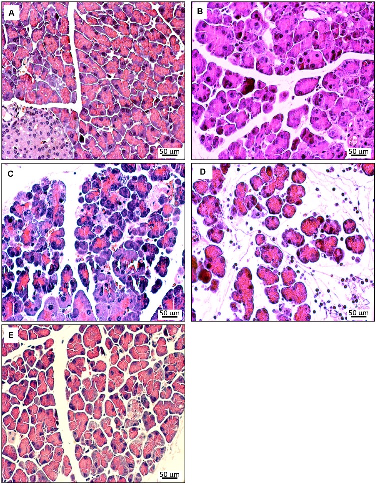
Figure 4. Histopathology of pancreatic lesions in cerulein-treated 12 months old UCP2-/- mice.
Pancreatic sections were stained with H&E. Photographs A–E display representative examples of pancreatic damage at the time points 0 h, 3 h, 8 h, 24 h and 7 days after the start of cerulein treatment. In addition to the findings described in Fig. 3, more pronounced histopathological changes at the time points 24 h (D – edema and presence of interstitial inflammatory cells) and day 7 (E – edema and cell damage) were observed.

