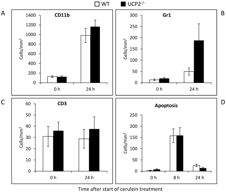
Figure 6. Immunohistochemical analysis of leucocyte infiltrates and quantification of apoptotic cells in cerulein-treated WT and UCP2-/- mice.
Pancreatic sections of 12 months old mice at the indicated time points after initiation of cerulein treatment (n = 8 per group) were processed applying the ABC technique and antibodies to CD11b (A), Gr1 (B) and CD3 (C), or stained with the ApopTag Kit for the detection of apoptotic cells (D). Positive-stained cells were counted, and mean values ± SEM were calculated. *P<0.05 (WT versus corresponding UCP2-/- mice).

