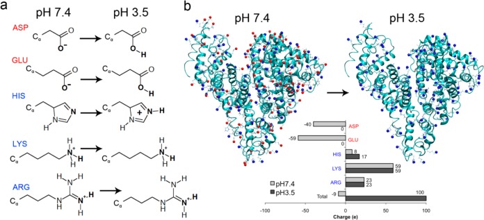
Figure 1.

Determination of ionization state of titratable residues for simulations. (a) Chemical structures of titratable residues (ASP, GLU, HIS, LYS, ARG) and the resulting ionized structure at pH 3.5. Red and blue color denotes negatively and positively charged residues, respectively. (b) Localization of ionized residues on albumin at pH 7.4 and pH 3.5. Inset shows the total charge per residue type and for the protein overall at the two pH values. At pH 7.4, the protein has a total charge of −9, while at pH 3.5 the charge is +100 (including the amine terminal group).
