
Figure 5.
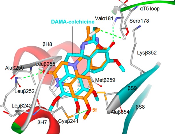
Predicted binding mode of 5f (orange stick) with tubulin (PDB code: 1SA0) and overlapping with DAMA-colchicine (cyan, the bound ligand of 1SA0). Surrounding amino acid side chains are shown in gray stick format and are labeled. Hydrogen bonds are shown by green dashed lines, and the distance between ligands and protein is less than 3 Å.